Treatment for melanoma in children
The incidence of melanoma, one of the most harmful forms of skin cancer, is increasing among children and teenagers. In fact, melanoma rates in the United States doubled from 1982 to 2011 and have continued to rise.
While melanoma represents only about 5 percent of all skin cancers in the United States, it accounts for about 75 percent of all skin cancer deaths. If detected early, melanomas have a high cure rate. However, in an advanced stage, it can spread to other organs.
Melanoma’s unique presentation in children sets it apart from its appearance in adults. Childhood is crucial for developing moles, an important melanoma risk factor. Regular skin check-ups are recommended for children with a family or personal history of skin cancer.
If your child or a loved one is at increased risk of developing pediatric melanoma or experiencing unusual mole development, we are here to help.

Understanding Pediatric Melanoma
Melanoma is a severe form of skin cancer that begins in melanocytes, the cells that give our skin, hair and eyes their color. When these cells become cancerous, they can lead to melanoma.
To understand pediatric melanoma, it’s helpful to know a bit about how our skin works. Melanocytes are found in the deepest layer of our skin and play a key role in protecting against the sun’s ultraviolet (UV) rays. Although melanin offers some natural defense, UV exposure during childhood can still damage skin cells and increase melanoma risk.
One of the important aspects of a childhood melanoma diagnosis is the presence of one or more spots on the skin. Often considered harmless, they may be more serious if they exhibit irregular edges, color changes, or increased in size or shape.
Learning to tell the difference can help doctors find melanoma early and treat it successfully. Learning to tell the difference can help doctors find melanoma early and treat it successfully.
What melanoma looks like in children
Melanoma in children may appear as a new or changing mole, often larger than nearby moles. It may also look like a uniform pink, red, or flesh-colored bump rather than a dark spot.
Any fast-growing, unusual, or symptomatic lesion deserves prompt evaluation.
How serious is melanoma in children?
Melanoma in children is serious because it can spread quickly if not detected early.
With timely diagnosis and treatment, most early-stage melanomas have high survival rates. Delayed care increases the risk of complications and more extensive treatment.
How melanoma in teens differs from younger children
Teens are more likely to develop melanoma that resembles adult patterns, which include darker, asymmetric lesions.
Younger children often present with amelanotic (less pigmented) or uniformly colored growths, which can be harder to recognize. These differences make professional skin exams especially important across age groups.
Childhood melanoma signs and symptoms
When recognizing melanoma in children, it’s essential to be aware of the common signs and symptoms. Melanoma in children might not always look the same as in adults, making early detection even more crucial.
Common signs and symptoms include:
- Unusual moles that may be larger, have irregular edges or have multiple colors
- An existing mole that changes in size, shape or color or begins to bleed
- New moles that appear after age four
- Any mole or spot that itches, bleeds or hurts
- A sore that does not heal within a few weeks
- Raised lumps or bumps on the skin’s surface
- Dark lines on nails, hands, feet or in the mouth
Identifying the early warning signs of melanoma is essential and knowing when to worry about a mole on a child is crucial for early diagnosis and successful treatment.
Pediatric melanoma can be more challenging to detect, but being vigilant and examining any unusual skin conditions or changes promptly is the best way to ensure a child’s health and well-being.
ABCDE signs adapted for pediatric melanoma
Melanoma in children can look different from adult melanoma. While the standard ABCDEs still matter, dermatologists also watch for additional patterns that are more common in younger patients:
- A – amelanotic or atypical
Children’s melanomas are more likely to be pink, red, or skin-colored, not the classic dark brown or black. - B – Bump
Instead of flat moles, pediatric melanomas often appear as a new raised bump or nodule that keeps growing. - C – Color uniformity (not variety)
Unlike adults, children may have lesions that are one solid color, even though they’re concerning. Any new, uniform-colored growth that changes quickly should be checked. - D – De novo (new)
Many pediatric melanomas occur in areas where no mole previously existed. A brand-new growth that looks unusual or changes rapidly should be evaluated right away. - E – Evolution
Any change (e.g., growth, firmness, bleeding, itching, or a shift in color or shape) is one of the most important warning signs in kids.
Children often present with “the ugly duckling sign,” meaning the spot looks noticeably different from their other moles, even if it doesn’t fit the classic ABCDE signs.
Symptoms that require urgent evaluation
- A new bump or growth that enlarges quickly
- A mole or spot that begins to bleed, ooze, or form a crust
- Sudden color change, especially if the lesion becomes red, pink, or skin-colored
- A mole that becomes painful, tender, or itchy
- A spot that looks noticeably different from the child’s other moles (“ugly duckling sign”)
- A lesion that becomes firm, raised, or starts to protrude from the skin
- A mole with redness or swelling around it
- A sore that doesn’t heal
- Pigment spreading beyond the borders of a mole
- Any new growth in a child with a personal or family history of atypical moles or melanoma
Causes and risk factors for pediatric melanoma
Risk factors for pediatric melanoma
Understanding the risk factors associated with pediatric melanoma is essential in protecting your child’s skin health and overall well-being.
Common risk factors include:
- A family history of melanoma or other skin cancers
- Prolonged exposure to harmful ultraviolet (UV) rays from the sun
- Living in sunny regions or spending extended periods outdoors without proper sun protection
- Fair skin, which is more susceptible to UV radiation
- Numerous or atypical moles
- Severe sunburns during childhood
- Conditions or treatments that weaken the immune system
- A personal history of skin cancer
- Tanning bed use
Genetic factors and family history
Some children inherit genetic traits that increase their risk for melanoma. Certain genetic syndromes can also increase mole development.
Families with a strong melanoma history may benefit from routine dermatologist-led skin checks.
Sun exposure and UV damage in childhood
Childhood is a crucial period for UV damage because young skin is more sensitive and therefore more vulnerable to damage. Even a few blistering sunburns can significantly increase melanoma risk later in life.
Consistent sun protection is one of the most effective prevention strategies. The expert dermatologists at Advanced Dermatology, P.C., recommend wearing a daily sunscreen of SPF 30 or higher.
Large or atypical moles in children
Children with large, congenital, or atypical moles have a higher risk of melanoma. These moles may require long-term monitoring to track changes.
Dermatologists may recommend routine photography or targeted exams to ensure early detection.
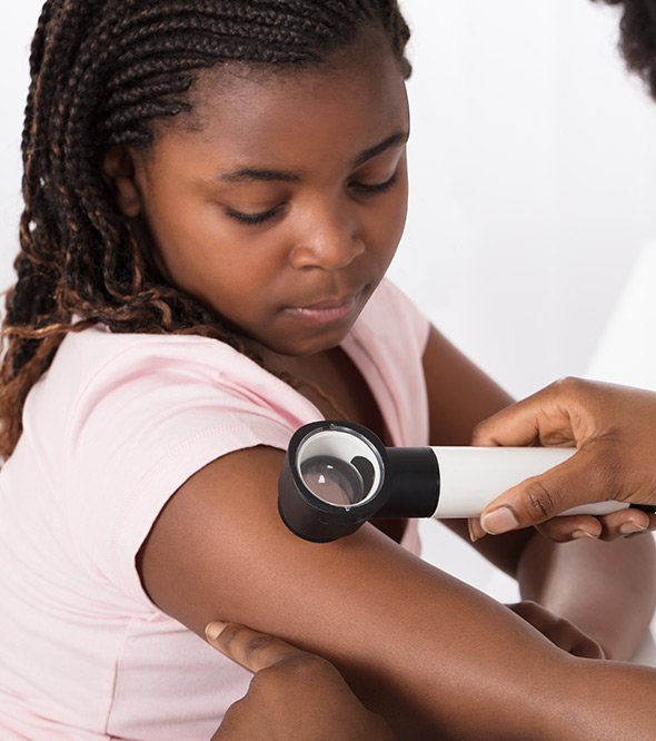
How pediatric melanoma is diagnosed
Diagnosing childhood skin cancer
Biopsy is a crucial step in diagnosing pediatric skin conditions, including melanoma. It allows doctors to examine the removed tissue under a microscope to determine if it’s cancerous.
For children, biopsy is the gold standard in confirming a melanoma diagnosis.
A child may receive four main types of biopsies for a suspected melanoma diagnosis:
- Excisional Biopsy
The entire mole or suspicious area is removed in this procedure. It’s the most common method for diagnosing melanoma. The tissue is then sent to a laboratory for analysis. - Incisional Biopsy
An incisional biopsy may be performed if a mole is too large to remove entirely. In this case, only a portion of the mole is removed for examination. - Punch Biopsy
A punch biopsy tool removes a small, circular section of the mole or skin lesion. This method is suitable for smaller moles - Shave Biopsy
A shave biopsy may be performed for surface-level moles. A scalpel shaves off the top layers of the mole for further examination.
During the biopsy, our dermatologists assess the type of melanoma, its depth and whether it has spread. This information guides the treatment plan, ensuring the best approach to care.
Pediatric dermatologist skin exam
A pediatric skin exam includes a careful evaluation of all moles and lesions, including those on the scalp, nails, and soles of the feet. Dermatologists look for subtle changes that can be easily missed at home.
These exams help establish a baseline for future comparison.
Melanoma biopsy in children
Biopsies in children are performed with gentle techniques to ensure comfort and accuracy. Local anesthesia is typically used so the child does not feel pain during the procedure. A dermapathologist then analyzes the sample.
When imaging tests are used
Imaging tests may be ordered if the melanoma is deeper or if there is concern about spread. Ultrasound, CT scans or MRI may help evaluate nearby lymph nodes or internal organs.
These tests support more precise staging and treatment planning.
Treatment options for childhood melanoma
In cases of early-stage melanoma, our experienced dermatologists often perform a specialized procedure known as an excision. During this procedure, they remove the melanoma and a margin of healthy tissue surrounding it to ensure all cancerous cells are taken out. This step minimizes the chance of the cancer returning. When the melanoma is small and localized, excision can offer a high cure rate.
In more advanced cases of melanoma, where the cancer may have spread to other parts of the body, specialized treatment is necessary. Our team, dedicated to your child’s well-being, will refer you to a specialist who is an expert in treating advanced melanoma.
These specialists are often oncologists, surgeons or other medical professionals with extensive experience managing complex pediatric melanoma cases.
Depending on the specific circumstances, they will work closely with you to create a tailored treatment plan that may include surgery, chemotherapy, immunotherapy, targeted therapy or radiation.
Pediatric melanoma surgery
Surgery is the main treatment for early-stage melanoma.
Dermatologists or surgeons remove the tumor along with surrounding tissue to ensure clear margins. Most children recover quickly and resume normal activities soon after.
Sentinel lymph node evaluation
A sentinel lymph node biopsy may be recommended if the melanoma is deeper or shows aggressive features. This test helps determine whether cancer cells have spread to nearby lymph nodes.
Immunotherapy and targeted therapy
For advanced melanoma, immunotherapy and targeted therapy may help the immune system recognize and attack cancer cells. These treatments are carefully selected based on the tumor’s characteristics. Pediatric oncologists monitor children closely to ensure safe, effective care.
Coordination with pediatric oncology
When melanoma is advanced or requires specialized therapies, dermatologists collaborate with pediatric oncologists. This team-based approach ensures comprehensive support from diagnosis through treatment.
Preventing melanoma in children
Shielding your child from the risk of melanoma begins with taking proactive measures.
Sun safety practices are crucial in preventing skin cancer, especially in children.
To reduce your child’s risk of developing pediatric melanoma, you should:
- Use SPF 30+ sunscreen daily
- Encourage hats, long sleeves and sunglasses
- Avoid tanning beds
- Monitor moles regularly
- Seek dermatology care for changing lesions
- Choose lightweight protective clothing
- Educate about sun safety
- Use shade when outdoors
- Reapply sunscreen as needed
- Avoid peak sun hours when possible (10:00 a.m. – 4:00 p.m.)
These measures can significantly reduce the risk of pediatric melanoma and promote overall skin health in children.
Sun protection and daily skin safety
Daily sunscreen use, protective clothing and shade can significantly reduce UV exposure. Children should apply sunscreen 15 minutes before going outside and reapply every two hours.
These simple habits can help prevent long-term skin damage.
Monitoring childhood moles over time
Regular mole checks help track changes that may signal melanoma.
Dermatologists often recommend annual exams for high-risk children. Parents can also use photos to monitor subtle changes between visits.
Reducing UV exposure for high-risk children
High-risk children benefit from stricter sun protection, including wearing UPF clothing and avoiding midday outdoor activities. Families should also be cautious during vacations, sports or other activities that involve prolonged sun exposure. Extra prevention helps offset inherited or medical risk factors.
Timely and complete care for melanoma in children
When it comes to your child’s health, we prioritize offering the most effective, comprehensive care.
Our experienced team is here to provide comprehensive treatment for all your medical dermatology needs. If your family is facing a pediatric melanoma diagnosis, rest assured that we’re here to guide you every step of the way.
Your child’s well-being is our top concern, and we’re committed to providing the best care possible.
Benefits of early diagnosis and treatment
When melanoma is found early, less invasive procedures are typically needed. Early diagnosis can also increase the likelihood of successful treatment, reduce the risk of spread, and support a full recovery.
Why choose advanced dermatology for pediatric melanoma
At Advanced Dermatology, P.C., your child receives care from experienced dermatologists trained in identifying, diagnosing and treating pediatric melanoma.
Our team provides thorough evaluations and personalized treatment plans.
Frequently Asked Questions
Is melanoma common in children?
Melanoma is rare in children but does occur, especially in those with genetic risk factors or significant sun exposure. Early evaluation helps ensure timely diagnosis and treatment.
How fast does melanoma grow in children?
Growth rates vary. Some pediatric melanomas grow quickly, making early detection critical.
What causes melanoma in children?
Causes include genetic predisposition, UV exposure, and the presence of atypical or congenital moles.
Can childhood moles turn into melanoma?
Most moles are harmless, but some atypical or congenital moles carry a higher risk and should be monitored.
When should a child’s mole be checked by a dermatologist?
Any changing, symptomatic, or unusual mole should be evaluated promptly. Children with higher risk should have routine dermatologist-led exams. Watch for any changes in growth, firmness, bleeding, itching, or a shift in color or shape.